Hilton’s Method: A Detailed Guide to Deep Abscess Drainage
This blog post provides a comprehensive overview of Hilton’s method, a surgical technique used to drain deep abscesses. We’ll explore its indications, contraindications, step-by-step procedure, post-operative care, and potential complications. Understanding this procedure is crucial for healthcare professionals and patients alike.
Understanding Hilton’s Method
Hilton’s method is a minimally invasive surgical technique for draining deep abscesses. Unlike traditional incision and drainage (I&D), which involves a wider incision, Hilton’s method utilizes blunt dissection through a small incision. This approach minimizes damage to surrounding vital structures like nerves, blood vessels, and important anatomical landmarks. The technique is particularly valuable when dealing with abscesses in sensitive areas like the neck, axilla, perineum, and thigh.
When is Hilton’s Method Appropriate?
Hilton’s method is the preferred approach when:
- Deep-seated abscesses are present: The abscess is located deep within the tissues, making direct incision risky.
- Proximity to vital structures: The abscess is near major nerves, arteries, or veins, necessitating a gentler approach to avoid iatrogenic injury.
- Blunt dissection is safer: The anatomical location or patient condition makes blunt dissection a safer alternative to sharp dissection.
When is Hilton’s Method NOT Appropriate?
Hilton’s method is contraindicated in:
- Superficial abscesses: For superficial abscesses, standard I&D is sufficient and less invasive.
- Unfit patients: Patients who are medically unfit for minor surgical procedures should not undergo this procedure without a thorough evaluation.
Essential Instruments and Preparation
Performing Hilton’s method requires a sterile surgical field and the following instruments:
- Sterile gloves and drapes
- Scalpel (typically a #11 or #15 blade)
- Artery forceps (Kelly or Mosquito forceps)
- Blunt-tipped scissors (Mayo scissors)
- Suction apparatus (optional, but highly recommended)
- Normal saline for irrigation
- Corrugated rubber or Penrose drain
- Antiseptic solution (povidone-iodine or chlorhexidine)
- Local anesthetic (1% lidocaine without epinephrine)
- Sterile gauze and dressing material
Prior to the procedure, informed consent must be obtained from the patient. The surgical site should be meticulously cleaned and draped using sterile technique. Local anesthesia, typically 1% lidocaine, is injected around the abscess, avoiding direct injection into the purulent material.
Step-by-Step Procedure: A Detailed Guide
The procedure involves several key steps:
1. Incision
A small stab incision (approximately 1.5-2 cm) is made at the point of maximum fluctuance (the area where the abscess feels softest) or in a dependent position to ensure optimal drainage. The incision should ideally follow Langer’s lines to minimize scarring.
2. Blunt Dissection: The Core of Hilton’s Method
Closed artery forceps or blunt-tipped scissors are inserted into the incision. The instruments are slowly advanced through the subcutaneous tissue, gently opening their jaws to separate tissue layers bluntly. This minimizes the risk of injuring blood vessels or nerves. The procedure continues until the abscess cavity is reached, indicated by a sudden release of pus under pressure.
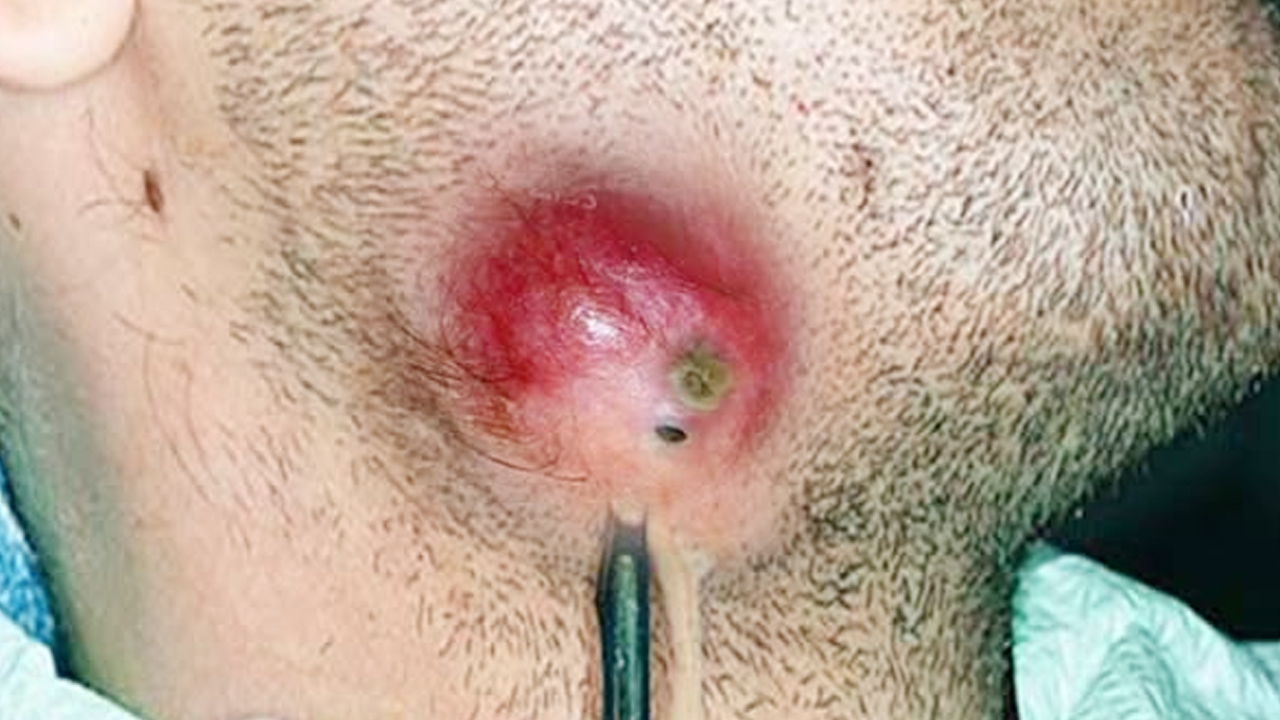
3. Drainage and Exploration
Pus is allowed to drain fully, or suction may be used if the abscess is under significant pressure. The abscess cavity is gently probed with forceps to break up any loculi (pus-filled pockets). Irrigation with normal saline is performed, and a sample of pus should be sent for microbiological culture and sensitivity testing.
4. Drain Placement
A corrugated or Penrose drain is inserted to prevent premature closure of the abscess cavity and ensure continued drainage. The drain is secured with sutures or dressings.
5. Dressing Application
A sterile, absorbent dressing is applied to the wound, which is then labeled and monitored for continued drainage.
Post-Operative Care and Medical Management
Post-operative care is crucial for successful healing and preventing complications:
Antibiotic Therapy
Antibiotic therapy may or may not be necessary, depending on the severity of infection and the patient’s clinical presentation. Empiric antibiotic treatment may be initiated if there are signs of systemic infection or cellulitis. The choice of antibiotic is guided by local antibiotic susceptibility patterns and the results of pus culture.
Pain Management
Analgesics, such as ibuprofen or paracetamol, are typically prescribed for pain management. Opioids may be considered for short-term use if pain is severe.
Wound Care
Daily dressing changes are necessary until the wound heals completely. The drain is typically removed after 48-72 hours, or when drainage ceases.
Follow-Up and Potential Complications
Regular follow-up is essential to monitor healing and detect any complications. Patients should be instructed to report any worsening pain, increased redness, swelling, fever, or reaccumulation of pus. Referral or readmission may be necessary if complications such as spreading cellulitis, deep space infections (e.g., Ludwig’s angina), or necrotizing fasciitis arise.
Clinical Example
A 27-year-old male presented with a 3-day history of painful axillary swelling. He was afebrile with a tender, fluctuant swelling. Hilton’s method was performed under local anesthesia. Pus was sent for culture, revealing methicillin-sensitive Staphylococcus aureus (MSSA). He was treated with incision and drainage (I&D) and amoxicillin-clavulanate. He recovered well, and the drain was removed after 2 days.
This blog post provides a comprehensive overview of Hilton’s method. However, this information is for educational purposes only and should not be considered medical advice. Always consult with a qualified healthcare professional for any health concerns.