Neck Cysts: When They Squirt and Ooze
A sudden squirting or oozing of fluid from a neck cyst is a dramatic event, usually signifying one of two scenarios: the cyst has ruptured spontaneously, or a healthcare professional has intentionally drained it. Let’s delve into the details of this concerning, yet relatively common, occurrence.
Understanding Neck Cysts and Their Contents
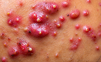
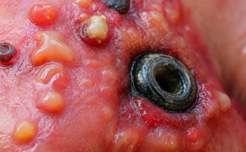
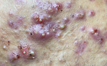
A cyst is essentially a sac filled with various substances. In the neck, the most prevalent types include epidermoid cysts (filled with keratin and sebum, giving a characteristic cheese-like consistency), branchial cleft cysts (congenital and often near the sternocleidomastoid muscle), and infected lymph nodes or abscesses (containing pus). The expulsion of cyst contents – that “squirting” or “oozing” – is a direct result of built-up internal pressure, forcing a release either through spontaneous rupture or via medical intervention (incision and drainage, or I&D).

The Telltale Signs: Deciphering the Fluid
The appearance of the released fluid provides valuable diagnostic clues. A thick, cheesy substance strongly suggests an epidermoid cyst. Yellowish-green, foul-smelling pus points towards infection or an abscess. Clear or slightly bloody fluid might indicate a lymphatic cyst or a ruptured cyst wall. A precise diagnosis often requires laboratory analysis.
Clinical Management: From Drainage to Excision
Managing a squirting or oozing neck cyst involves several key steps.
Incision and Drainage (I&D) Procedure
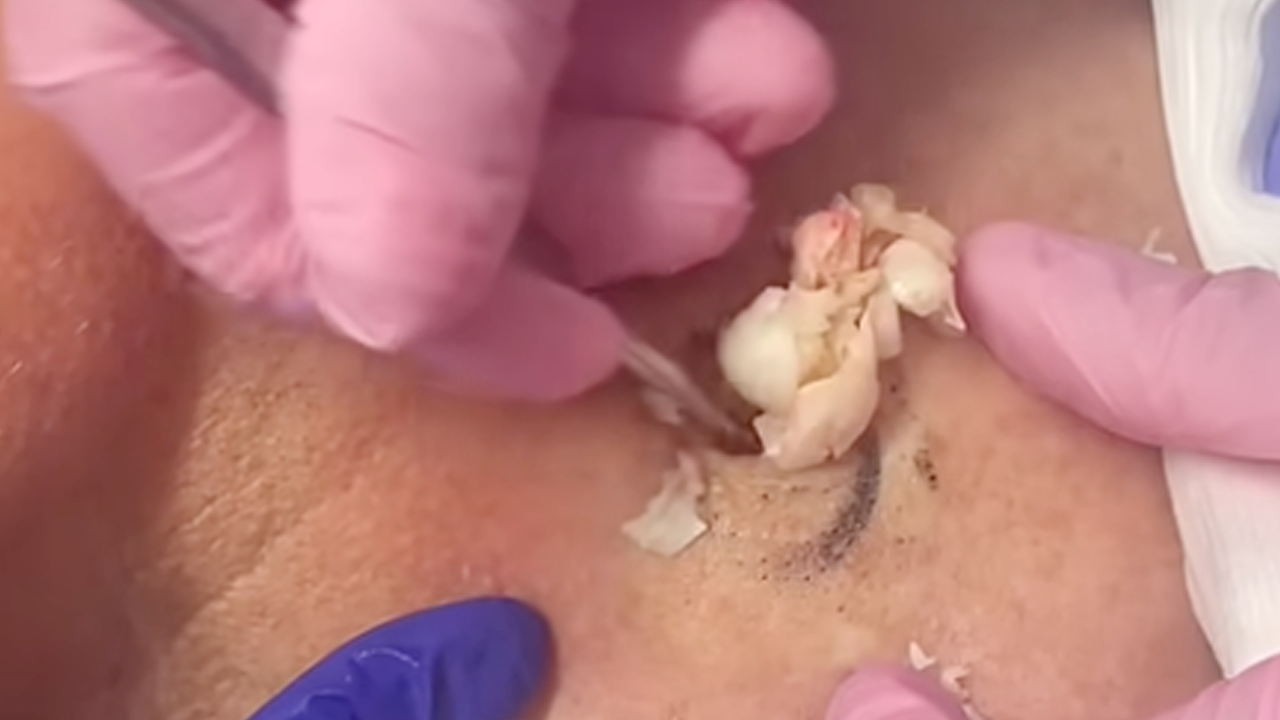
This procedure, performed under local anesthesia, involves a small incision made at the cyst’s most prominent point. Gentle pressure or sterile instruments are then used to express the contents. If the pressure is high, the fluid may indeed squirt out. The cavity is subsequently rinsed with saline or antiseptic solution, and a temporary drain or wick might be inserted.
Culture, Sensitivity Testing, and Antibiotics
If infection is suspected (as indicated by pus), a sample is sent for culture and sensitivity testing to identify the responsible bacteria and determine the most effective antibiotic. Empirical antibiotic treatment (such as clindamycin or amoxicillin-clavulanate) is often initiated immediately, later adjusted based on lab results.

Definitive Treatment: Complete Excision
Once the inflammation subsides, the optimal course of action is to surgically remove the entire cyst capsule. This prevents recurrence, a common complication if only the contents are drained and the cyst wall remains.
Potential Complications and Post-Drainage Care
While I&D and antibiotic treatment effectively address immediate concerns, potential complications necessitate vigilance. Recurrence is a significant risk if the cyst wall isn’t fully excised. Secondary infection, scarring, sinus tract formation, and the development of abscesses are also possibilities, particularly concerning in the delicate neck region. Severe complications could include cellulitis or deeper neck space infections.
If the cyst has already been drained, maintaining a clean and dry environment is crucial. Antibiotic ointment may be recommended. Closely monitor for any signs of infection (increased redness, swelling, fever) and seek immediate medical attention if they arise. Crucially, avoid squeezing or manipulating the area.

Forehead Biopsies for Squamous Cell Carcinoma (SCC): A Modified Approach
Diagnosing squamous cell carcinoma (SCC) on the forehead requires a tailored approach due to the unique anatomical and cosmetic considerations of this area. Standard biopsy techniques must be modified to balance diagnostic accuracy with optimal cosmetic outcomes.
Why Modify Standard Biopsy Techniques?
Standard shave, punch, and excisional biopsies are widely used for SCC diagnosis. However, on the forehead, modifications are vital to:
- Preserve cosmetic appearance: Minimizing scarring is paramount on the visible forehead.
- Avoid damage to underlying structures: The forehead’s proximity to vital structures requires careful planning.
- Ensure complete and accurate sampling: Adequate tissue must be obtained for an accurate diagnosis.
The choice of technique hinges on factors like lesion size, depth, and the clinician’s assessment of potential invasion.
Modified Biopsy Techniques for Forehead SCC
Various modified techniques are adapted for forehead SCC, each with specific indications and considerations:
1. Modified Shave Biopsy
- Indications: Superficial, raised lesions.
- Technique: A scalpel or razor blade removes the lesion and a portion of the underlying dermis. Hemostasis is achieved via pressure, chemical agents, or electrocautery. Sutures are usually unnecessary.
- Advantages: Minimally invasive, quick, and cosmetically favorable.
- Considerations: May not provide a full-thickness sample, potentially missing deeper invasion.
2. Modified Punch Biopsy
- Indications: Lesions of uncertain depth or when full-thickness sampling is crucial.
- Technique: A circular blade extracts a cylindrical tissue sample, including epidermis, dermis, and superficial subcutaneous fat. Sutures may or may not be required.
- Advantages: Provides a full-thickness sample, suitable for invasive SCC.
- Considerations: May leave a small scar if sutures are used.
3. Modified Excisional Biopsy
- Indications: Small lesions where complete removal is both feasible and desired.
- Technique: An elliptical incision removes the lesion and a margin of surrounding skin. The wound is then closed with sutures.
- Advantages: Allows complete histological assessment of the lesion and margins.
- Considerations: May result in a more noticeable scar; careful planning to align with skin tension lines is essential for optimal cosmetic results.
Post-Biopsy Care: Ensuring Proper Healing
Regardless of the technique used, post-biopsy care is crucial. Maintaining a clean and dry environment, following wound care instructions, and monitoring for signs of infection are essential. Regular follow-up appointments are necessary to discuss pathology results and subsequent management.